All Images
News Release 04-076
Staying on the Path – One Atom at a Time
New percolation model may allow researchers to study biochemistry at the atomic level
This material is available primarily for archival purposes. Telephone numbers or other contact information may be out of date; please see current contact information at media contacts.

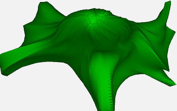
Surface rendering of a neuron. A Delauney tessellation scheme was used to discretize the volume. This new approach can be applied to any vertically single-branched cell.
Credit: Yun-Bo Yi and Ann Marie Sastry, University of Michigan
Download the high-resolution TIFF version of the image. (996 KB)
Use your mouse to right-click (Mac users may need to Ctrl-click) the link above and choose the option that will save the file or target to your computer.

Rendering of the interior of a 3-D model of a neuron. 200 confocal microscope images (each only 0.1mm in thickness) were used to generate the image. The smallest resolvable feature (a single pixel in the image) is about 0.1mm in size.
Credit: Yun-Bo Yi and Ann Marie Sastry, University of Michigan
Download the high-resolution TIFF version of the image. (649 KB)
Use your mouse to right-click (Mac users may need to Ctrl-click) the link above and choose the option that will save the file or target to your computer.

Clusters of two, three and four permeable ellipsoids, generated from the percolation simulations of Yun-Bo Yi and Ann Marie Sastry.
Credit: Yun-Bo Yi and Ann Marie Sastry, University of Michigan
Download the high-resolution TIFF version of the image. (625 KB)
Use your mouse to right-click (Mac users may need to Ctrl-click) the link above and choose the option that will save the file or target to your computer.
